Field Tested & Hunter Approved
Labrador Health

At Guns Up Labradors our priority is to produce healthy puppies that are structurally and genetically sound, and demonstrate the true Labrador Retriever temperament which make the breed such wonderful family members.
We also strive to maintain the hunting instinct/drive that Labradors Retrievers are known for, and is why they are such exceptional gun dogs and working companions.
Breeding sound, quality Labrador Retrievers is a costly and time-consuming undertaking if done properly. As responsible/ethical breeders we plan each litter carefully, maintain our high standards and strive to achieve improvement with each generation.
We do not breed on pre-liminary OFA Hip and Elbow clearances. We only choose dogs who have passed their final OFA clearances at 2 two years of age to participate in our very limited breeding program.
Anyone interested in owning or breeding a Labrador Retriever should become familiar with the various medical conditions, and appropriate health clearances, before deciding to obtain a Labrador.
The following are known health issues in the Labrador Retriever breed:

Hip Dysplasia
Hip Dysplasia typically develops because of an abnormally developed hip joint, but can also be caused by cartilage damage from a traumatic fracture. With cartilage damage or a hip joint that isn’t formed properly, over time the existing cartilage will lose its thickness and elasticity. This breakdown of the cartilage will eventually result in pain with any joint movement. No one can predict when or even if a dysplastic dog will start showing clinical signs of lameness due to pain. Severity of the disease can be affected by environmental factors, such as caloric intake or level of exercise. There are a number of dysplastic dogs with severe arthritis that run, jump, and play as if nothing is wrong and some dogs with barely any arthritic x-ray evidence that are severely lame.
- Visit OFA.org to learn more about Hip Dysplasia including: Screening procedure, grade classifications and treatment options.
- Visit What is PennHip? to learn more about the PennHip screening method.

Elbow Dysplasia
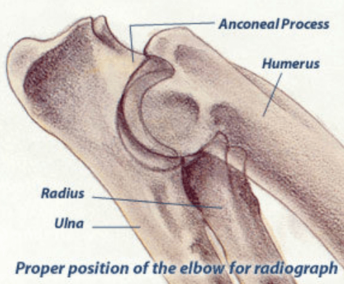
Elbow dysplasia is a general term used to identify an inherited polygenic disease in the elbow. Three specific etiologies make up this disease and they can occur independently or in conjunction with one another. These etiologies include:
- Pathology involving the medial coronoid of the ulna (FCP)
- Osteochondritis of the medial humeral condyle in the elbow joint (OCD)
- Ununited anconeal process (UAP)
Studies have shown the inherited polygenic traits causing these etiologies are independent of one another. Clinical signs involve lameness which may remain subtle for long periods of time. No one can predict at what age lameness will occur in a dog due to a large number of genetic and environmental factors such as degree of severity of changes, rate of weight gain, amount of exercise, etc. Subtle changes in gait may be characterized by excessive inward deviation of the paw which raises the outside of the paw so that it receives less weight and distributes more mechanical weight on the outside (lateral) aspect of the elbow joint away from the lesions located on the inside of the joint. Range of motion in the elbow is also decreased.
- Visit OFA.org to learn more about Elbow Dysplasia including: Screening procedure, grade classifications and treatment options.
Companion Animal Eye Registry (CAER)
The purpose of the OFA Companion Animal Eye Registry (CAER) is to provide breeders with information regarding canine eye diseases so that they may make informed breeding decisions in an effort to produce healthier dogs. CAER certifications will be performed by Board Certified (ACVO) Veterinary Ophthalmologists.
For Labrador Retrievers, the examinations are generally conducted at 7 weeks of age, then annually up to the age of 10 years.
The procedure, which is conducted yearly, involves a careful and comprehensive examination of the eye. To start with, the dog’s pupils are dilated with eye drops. The examiner then illuminates the eye with a penlight to look for any key abnormality.
The eye is then examined in detail using a slit lamp bio-microscope to identify any diminutive anomalies in the lens, cornea, and in the anterior chamber. During this part of the exam anomalies such as distichia, cataracts, vitreal degenerations, and corneal dystrophy may be noticed.
Lastly, the retina is examined using an ophthalmoscope (usually an indirect ophthalmoscope). This exam provides the examiner a lucid view of all the parts of the retina. The indirect ophthalmoscope device offers the veterinarian with proper optics and a light source. Problems such as Progressive Retinal Atrophy, Retinal Dysplasia, optic nerve hypoplasia, choroidal hypoplasia may be revealed during this part of the examination.
If any problems are identified during these examinations, they are recorded in an official form by the Ophthalmologist. Regardless of whether owners submit their CAER exam forms to the OFA for “certification,” all CAER exam data is collected for aggregate statistical purposes to provide information on trends in eye disease and breed susceptibility. Clinicians and students of ophthalmology as well as interested breed clubs, individual breeders and owners of specific breeds will find this useful.

Progressive Retinal Atrophy (prcd-PRA)

The OptiGen prcd-PRA test is a DNA-based test that helps you avoid one form of Progressive Retinal Atrophy (PRA). PRA refers to a group of diseases that cause the retina of the eye to degenerate slowly over time. The result is declining vision and eventual blindness. “prcd” stands for “progressive rod-cone degeneration” which is the type of PRA known in several breeds.
AFTER reading the information on this page, you can link to information specifically about the breed in which you are interested.
- Visit Optigen for more information about Progressive Retinal Atrophy (prcd-PRA) including testing.
Retinal Dysplasia/Ocuskeletal Dysplasia (RD/OSD)
Retinal Dysplasia-retinal folds (RD) is a common clinical observation in many dog breeds. Since many retinal folds are benign and of unknown heritability, veterinary ophthalmologists will often advise that breeding dogs with RD is an acceptable option. However, in Labrador Retrievers RD is of much greater concern. RD in Labradors will cause a dog to fail a CAER examination, the recommended annual eye examination that is done in North America by certified veterinary ophthalmologists, diplomates of the American College of Veterinary Ophthalmology (ACVO).
In such cases, breeding is not advised because RD in these breeds can be an indication that the dog is a carrier of a serious inherited syndrome called OSD (OculoSkeletal Dysplasia). OSD is a severe condition in which the dogs show a variety of skeletal malformations, including shortened limbs (dwarfism), and blindness at an early age; the blindness results from a generalized malformation of the retina that causes a partial or full retinal detachment and cataracts.
- Visit Optigen for more information about Retinal Dysplasia/Ocuskeletal Dysplasia (RD/OSD) including testing.

Tricuspid Valve Dysplasia

ricuspid valve dysplasia (TVD) in dogs is a congenital heart condition In medical terms, dysplasia means a malformation; TVD is a condition in which the heart’s tricuspid valve forms improperly during embryonic development. Like the human heart, a dog’s heart is divided into two halves. The tricuspid valve is in the right half between the right atrium and the right ventricle. Its job is to prevent the back flow of oxygen-depleted blood as it flows from the right atrium to the right ventricle on its way to becoming oxygenated once again by the lungs via the pulmonary artery.
Made up of irregularly shaped flaps, the tricuspid valve forms a barrier when blood tries to work its way back into the right atrium as the right ventricle contracts. In utero, these flaps are adhered to the ventricle wall. Normally, cellular degeneration takes place and the flaps are detached. In dogs with TVD, this degeneration does not take place and the flaps remain connected to the ventricle wall, hampering the valve from doing its job of preventing the blood’s back flow. Blood is then regurgitated, or leaked, back into the right atrium, increasing the workload of the right side of the heart.
- Visit OFA.org to learn more about Cardiac Disease in dogs including: Screening procedures Auscultation and Echo Doppler, as well as grade classifications.
Exercise-Induced Collapse (EIC)
Exercise-Induced Collapse (EIC) is an inherited neuromuscular disorder affecting Labrador Retrievers.
EIC presents as exercise intolerance in apparently healthy dogs. Affected dogs are usually diagnosed before two years of age and appear normal during low to moderately strenuous activity. However, shortly after 5-20 minutes of strenuous exercise affected dogs will begin to walk with a wobbly, uncoordinated gait that often only affects the hind limbs. Dogs remain mentally alert and are not in pain during episodes of EIC.
In some circumstances, the symptoms of EIC can progress to full body weakness with low muscle tone (flaccid paralysis), confusion, loss of consciousness, seizures and very rarely, death. The episodes typically last 5-10 minutes and most dogs will completely recover within 15-30 minutes.
- Visit the University of Minnesota for more information about Exercise Induced Collapse (EIC) including testing.

Centronuclear Myopathy (CNM)

Centronuclear Myopathy is an inherited progressive muscle disease affecting Labrador retrievers.
Though the severity of symptoms is variable, affected dogs typically present between 6 weeks to 7 months of age with exercise intolerance, awkward gait and difficulty eating. As the disease progresses, symptoms also include generalized muscle Atrophy, downward flexion of the head and neck, low muscle tone and more frequent episodes of collapse when exposed to cold temperatures.
Progression of the disease tends to stabilize around one year of age and dogs typically have a normal life span, but affected dogs usually have life-long medical problems due to the underlying muscle disease.
- Visit CNM Online for more information about Centronuclear Myopathy (CNM) including: Symptoms, testing and care for a CNM affected dog.
Degenerative Myelopathy (DM)
Degenerative Myelopathy is an inherited neurologic disorder caused by a Mutation of the SOD1 gene known to be carried by Labrador retrievers. This mutation is found in many breeds of dog, though it is not clear for Labrador retrievers whether all dogs carrying two copies of the mutation will develop the disease. The variable presentation between breeds suggests that there are environmental or other genetic factors responsible for modifying disease expression.
The average age of onset for dogs with degenerative myelopathy is approximately nine years of age. The disease affects the White Matter tissue of the spinal cord and is considered the canine equivalent to amyotrophic lateral sclerosis (Lou Gehrig’s disease) found in humans. Affected dogs usually present in adulthood with gradual muscle Atrophy and loss of coordination typically beginning in the hind limbs due to degeneration of the nerves.
The condition is not typically painful for the dog, but will progress until the dog is no longer able to walk. The gait of dogs affected with degenerative myelopathy can be difficult to distinguish from the gait of dogs with hip dysplasia, arthritis of other joints of the hind limbs, or intervertebral disc disease. Late in the progression of disease, dogs may lose fecal and urinary continence and the forelimbs may be affected.
Affected dogs may fully lose the ability to walk 6 months to 2 years after the onset of symptoms. Affected medium to large breed dogs, such as the Labrador retriever, can be difficult to manage and owners often elect euthanasia when their dog can no longer support weight in the hind limbs.
- Visit Paw Print Genetics for more information about Degenerative Myelopathy including testing.

Skeletal Dysplasia Type 2

Several hereditary forms of disproportionate dwarfism or “short-legged” phenotype have been recognized by breeders of Labrador Retrievers over the years. A distinct form of a mild skeletal dysplasia type 2, termed SD2, is characterized by short legs with normal body length and width.
In contrast to more severe conditions such as Oculoskeletal Dysplasia (OSD) or radius curvus deformity, SD2 has a very subtle phenotype without any obvious ocular or auditory defects and without known secondary joint problems.
Still, this form of disproportionate dwarfism is not a desirable trait and can result in Labrador Retrievers failing to reach the height standard for the breed.
- Visit Optigen for more information about Skeletal Dysplasia Type 2 including testing.
Hereditary Nasal Parakeratosis (HNPK)
Hereditary Nasal Parakeratosis is an inherited disease affecting the nose of Labrador Retrievers. Beginning around 6 to 12 months of age, affected dogs develop dry, rough, gray to brown crusts and rarely, painful cracks on the tip of the nose. In some cases, lesions are also present on the haired area around the nose.
The noses of affected dogs are prone to superficial bacterial infections and often become depigmented over time. Affected dogs are otherwise healthy. Symptoms often wax and wane in severity over the dog’s life.
Though manageable, this disorder requires continuous topical therapy to prevent recurrence of excessive nasal crusting.
- Visit Paw Print Genetics for more information about Skeletal Hereditary Nasal Parakeratosis (HNPK) including testing.

Copper Toxicosis (Labrador Type)

Copper toxicosis (Labrador Retriever Type) is an inherited metabolic disease affecting dogs, resulting in chronic liver failure. Dogs with Copper Toxicosis have a decreased ability to excrete dietary copper from the body resulting in excessive copper storage in tissues and organs, including the liver, which can result in liver damage and subsequent cirrhosis.
Though the age of onset and speed of disease progression are variable, most affected dogs will present in middle age with non-specific signs of liver dysfunction including weight loss, lethargy, weakness, vomiting, diarrhea, and abdominal pain. In late stages of disease, affected dogs may develop signs of liver failure including abdominal swelling, jaundice, and neurological dysfunction. Dogs found to have one or two copies of the Mutation may benefit from certain therapies.
- Visit Paw Print Genetics for more information about Copper Toxicosis (Labrador Type) including testing.
Coat Color & D Locus Testing
DNA testing for coat color genotypes is available for many dogs breeds through several genetic testing laboratories, e.g., DDC Veterinary, Paw Print Genetics, VetGen, etc.
Breeders can use Coat Color DNA testing as a tool to more effectively manage mating patterns and make more informed breeding decisions. By identifying the color genotype of the parents, breeders can more accurately predict the colors of future progeny.
For Labrador Retrievers there are 3 accepted coat color phenotypes (that can be seen): Black, Yellow, and Chocolate.
There are 9 possible reported genotypes (that cannot be seen) for the E and B loci.
- Black Coat: 4 possible genotypes- EEBB (black with no hidden colors), EeBB (black with hidden yellow), EEBb (black with hidden chocolate), EeBb (black with hidden yellow and chocolate)
- Yellow Coat: 3 possible genotypes- eeBB (yellow with hidden black), eeBb (yellow with hidden black and chocolate), eebb (yellow with hidden chocolate- dogs will exhibit liver noses.
- Chocolate Coat: 2 possible genotypes- EEbb (chocolate with no hidden colors), Eebb(chocolate with hidden yellow)
D Locus (DNA marker tested – C.22G>A)
The D Locus is associated with the dilution or lightening effect of solid colors, with D being the dominant allele, the dd genotype results in the diluted effect (like grey, blue, silver, lilac, champagne).
The dd (Affected) or Dd (Carrier) genotypes are not natural, nor accepted in the Labrador Retriever breed. Diluted “colors” are a disqualification based on the published AKC and the LRC’s breed standard.
The dogs in our breeding program are tested free of the dilute genotypes. Their results are DD, meaning they Do Not carry the D Locus.
Please visit our “Designer Dogs Page” for more information about the Dilute gene, as well as other Designer breeds.
- Visit DDC Veterinary to read this complete article on Canine DNA Coat Color Testing and to learn more about Canine Coat Color Testing in general. Follow this link to view the DDC Coat Color Predictor Chart.


Information & Resources